Product Name
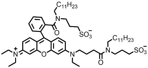
Cell membrane green fluorescent probe RMG3, 95%
Molecular Formula
C60H93N4O9S2-
Certificate of Analysis (COA)
RMG3 Cell Membrane Fluorescent Probe
Product Description
RMG3 is a cell membrane-specific fluorescent probe developed through structural modification of the Rhodamine B fluorophore. It enables rapid staining of live-cell plasma membranes without requiring washing steps while minimizing probe internalization.
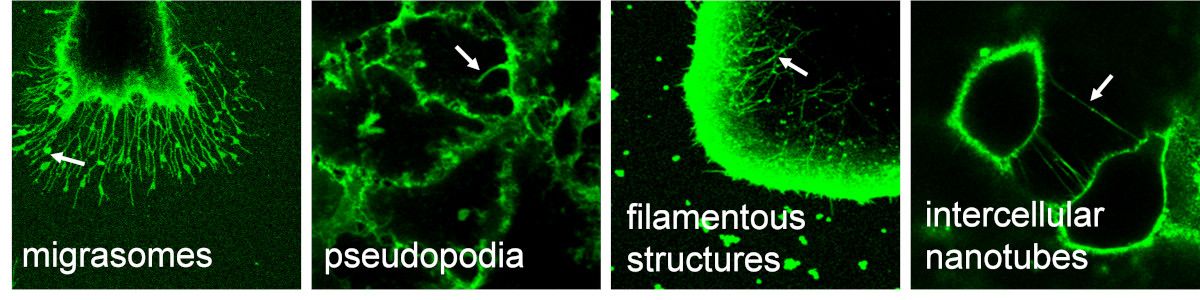
RMG3 exhibits excellent fluorescence brightness and provides high-resolution visualization of cellular membranes and fine membrane-associated structures, including filopodia, actin-rich protrusions, tunneling nanotubes (TNTs), migrasomes, and other membrane-derived structures. It serves as a reliable, convenient, and efficient tool for studying cell membrane morphology and dynamic membrane remodeling processes.
Excitation/Emission Wavelengths: 570/595 nm
Compatible Imaging Channels: FITC, TRITC, Cy3, and Texas Red filter sets.
Experimental Protocol
1. Preparation of RMG3 Working Solution
The product is supplied as a 1 mM stock solution.
Before use, dilute the stock solution with PBS or serum-free cell culture medium to a final concentration of 100 nM–2 μM. A working concentration of 1 μM is recommended for most applications.
Note:
Store the stock solution at −20°C or −80°C, protected from light. Aliquoting is recommended to avoid repeated freeze-thaw cycles.
2. Cell Staining
2.1 Suspension Cells (6-Well Plate)
a. Centrifuge suspension cells at 1000 rpm for 5 min at 4°C and discard the supernatant. Wash cells three times with PBS.
b. Add 1 mL of 1 μM RMG3 working solution and incubate at room temperature for 2–5 min.
Note: Incubation time may be optimized according to staining results.
c. Centrifuge at 1000 rpm for 5 min at 4°C and wash the cells with PBS 2–3 times.
d. Resuspend cells in serum-free culture medium or PBS and analyze by fluorescence microscopy or flow cytometry.
2.2 Adherent Cells
a. Seed cells onto sterile coverslips or culture dishes and allow them to adhere for 8–24 h before staining.
b. Remove the culture medium and wash cells three times with PBS.
c. Add 1 mL of 1 μM RMG3 working solution and incubate at room temperature for 2–5 min.
No washing step is required, and cells can be directly observed under a fluorescence microscope.
Note: For long-term imaging (>1 h), incubate cells with RMG3 for 2–5 min, wash three times with PBS, and replace with serum-containing culture medium to maintain cell viability and ensure optimal imaging results.
3. Fluorescence Imaging
The probe exhibits excitation and emission maxima at 570 nm and 595 nm, respectively.
Recommended laser lines include:
- 559 nm
- 561 nm
- 488 nm (compatible)
Compatible fluorescence filter sets include:
Precautions
- Optimize the concentration of RMG3 working solution according to the desired fluorescence intensity and experimental conditions.
- This product is intended for research use only and is not intended for therapeutic, diagnostic, household, or other applications.
- Appropriate personal protective equipment (PPE) should be worn during handling.
- In case of accidental contact with eyes or skin, rinse immediately with plenty of water and seek medical attention if necessary.
Frequently Asked Questions (FAQ)
1. What is RMG3 used for?
RMG3 is a live-cell membrane fluorescent probe specifically designed for rapid visualization of plasma membranes. It enables high-contrast imaging of cell membrane morphology and membrane-associated structures such as filopodia, tunneling nanotubes (TNTs), migrasomes, and membrane protrusions.
2. Does RMG3 require a washing step after staining?
In most live-cell imaging applications, washing is not required. After incubation with RMG3 for 2–5 minutes, cells can be directly imaged. For long-term observation (>1 hour), washing with PBS and replacing the staining solution with complete culture medium is recommended to maintain cell viability.
3. Can RMG3 be used for live-cell imaging?
Yes. RMG3 is specifically optimized for live-cell membrane staining. The probe rapidly labels cell membranes while minimizing intracellular internalization, making it highly suitable for dynamic live-cell imaging studies.
4. What is the recommended working concentration of RMG3?
The recommended working concentration is 1 μM. However, concentrations ranging from 100 nM to 2 μM may be used depending on cell type, imaging platform, and desired fluorescence intensity.
5. Which fluorescence channels are compatible with RMG3?
RMG3 has excitation and emission maxima at 570 nm and 595 nm, respectively. It is compatible with:
- FITC channel
- TRITC channel
- Cy3 channel
- Texas Red channel
Recommended laser lines include 559 nm and 561 nm.
6. Can RMG3 be used with flow cytometry?
Yes. In addition to fluorescence microscopy, RMG3 can be used for flow cytometric analysis of cell membrane labeling following the staining protocol for suspension cells.
7. Does RMG3 stain intracellular organelles?
RMG3 is designed to preferentially label the plasma membrane and exhibits minimal intracellular accumulation under recommended staining conditions. This helps achieve clear membrane-specific imaging with low background fluorescence.
8. Is RMG3 suitable for imaging fine membrane structures?
Yes. Owing to its high fluorescence brightness and membrane specificity, RMG3 provides excellent visualization of fine membrane structures, including:
- Filopodia
- Lamellipodia
- Tunneling nanotubes (TNTs)
- Migrasomes
- Cell-cell membrane connections
9. Can RMG3 be used with other fluorescent probes?
Yes. RMG3 can be combined with a variety of fluorescent dyes, fluorescent proteins, and organelle markers. However, users should verify spectral compatibility and optimize imaging parameters to minimize fluorescence overlap.
10. What cell types are compatible with RMG3?
RMG3 is suitable for a wide range of mammalian cells, including:
- Adherent cell lines
- Suspension cell lines
- Primary cells
- Stem cells
- Immune cells
Optimization of staining concentration and incubation time may be required for specific cell types.
11. Is RMG3 intended for clinical or diagnostic use?
No. RMG3 is intended for research use only (RUO) and is not approved for clinical, therapeutic, or diagnostic applications.